
RETINA & UVEA CARE
Macular Hole
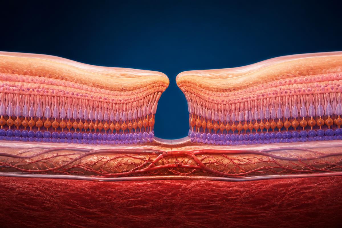
A macular hole is a small full-thickness opening at the centre of the retina. It commonly develops when age-related vitreous separation exerts traction on the fovea. Patients may notice central blur, distortion or a missing spot, while side vision usually remains unaffected.
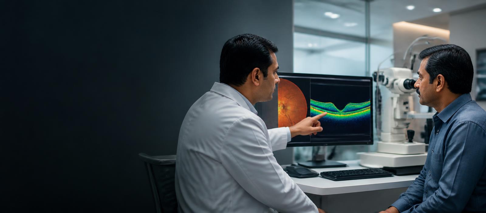
A macular hole differs from a retinal tear in the periphery. It affects the fovea, the point responsible for the sharpest central vision, and generally does not progress into a typical retinal detachment. OCT is essential because other conditions can mimic a hole on examination.
Vitrectomy releases vitreous traction and often includes peeling a very thin membrane from the macula. A gas bubble supports closure while the retina heals. The surgeon advises positioning based on hole size and technique; patients should not impose prolonged face-down positioning without specific instructions.
1. How a macular hole may present
Straight lines, faces or printed words appearing bent or interrupted
A blurred or missing central spot in one eye
Difficulty reading fine print or performing detailed tasks despite an updated spectacle prescription
2. Diagnosis and treatment pathway
Confirm with OCT
A high-resolution OCT scan shows the hole's width, depth and associated vitreous traction.
Decide between observation and surgery
Some small early holes may be monitored, but most full-thickness symptomatic holes are treated with vitrectomy and gas.
Support closure
After removal of traction, an internal limiting membrane peel and gas bubble help the edges flatten; positioning advice is individualised.
3. What patients should understand
Timing matters
Long-standing and larger holes may have lower closure and visual recovery potential than recent smaller holes.
The gas is temporary
Vision is very blurred while the bubble fills the eye and improves gradually as it absorbs.
Lens change is common
Cataract commonly progresses after vitrectomy in eyes that still have the natural lens.
4. Aftercare and risks
- Do not fly, travel to high altitude or receive nitrous-oxide anaesthesia until the gas bubble has fully disappeared
- Risks include infection, bleeding, raised pressure, retinal tear or detachment, cataract and failure of the hole to close
- Anatomical closure and visual improvement are not identical; final reading vision varies
Why choose Perfect Vision Laser Eye Hospital, Jaipur?
- Led by Dr. Virendra Agrawal, MD (Ophthalmology), AIIMS New Delhi, with a full-time specialist team
- Diagnosis-first approach with advanced imaging before any recommendation
- Comprehensive eye services available at one Jaipur centre
- Support for patients travelling from across Rajasthan and outside the state
- Cashless Mediclaim assistance, subject to your policy terms
Your specialists

Dr. Anju Bansal
Consultant — Vitreoretina, Uvea & ROP Specialist. MBBS, DNB (Ophthalmology), FVRS (Retina, Uvea & ROP) — Fellowship at Dr. Shroff's Charity Eye Hospital, New Delhi · Experience:
Every retinal condition requires a precise diagnosis and an individualised treatment strategy. My priority is to combine evidence-based care with compassion, clarity and continuity.
Dr. Virendra Agrawal
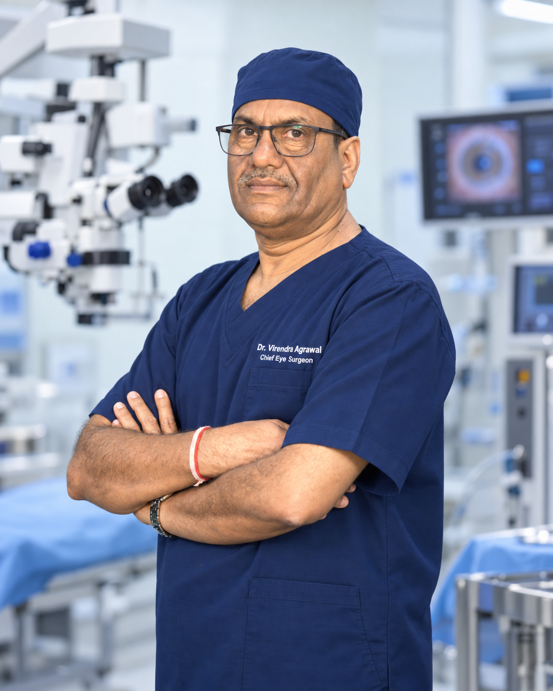
Medical Director & Chief Consultant — Cataract & Refractive Surgery. MD (Ophthalmology), DNB, MNAMS, FAICO (Cataract & Refractive Surgery) — Bodh Raj Sabarwal Gold Medalist (AIIMS, New Delhi) · Experience: 30+ Years of Ophthalmology
Every eye is different. The right procedure is the one that fits your cornea, your lens, your retina and the way you use your vision every day — not the one that is most advertised. Technology supports the decision; it never replaces the clinical evaluation.

Dr. Amit Mishra
Senior Consultant — Cataract, Refractive & Glaucoma. MBBS, MS (Ophthalmology) · Experience: 25+ Years of Ophthalmology
Advanced eye care is a balance of proven surgical technique and ongoing learning. Every patient deserves a careful evaluation, a clear explanation of options and a treatment plan built around their individual vision and eye health.
Related technology
Retina
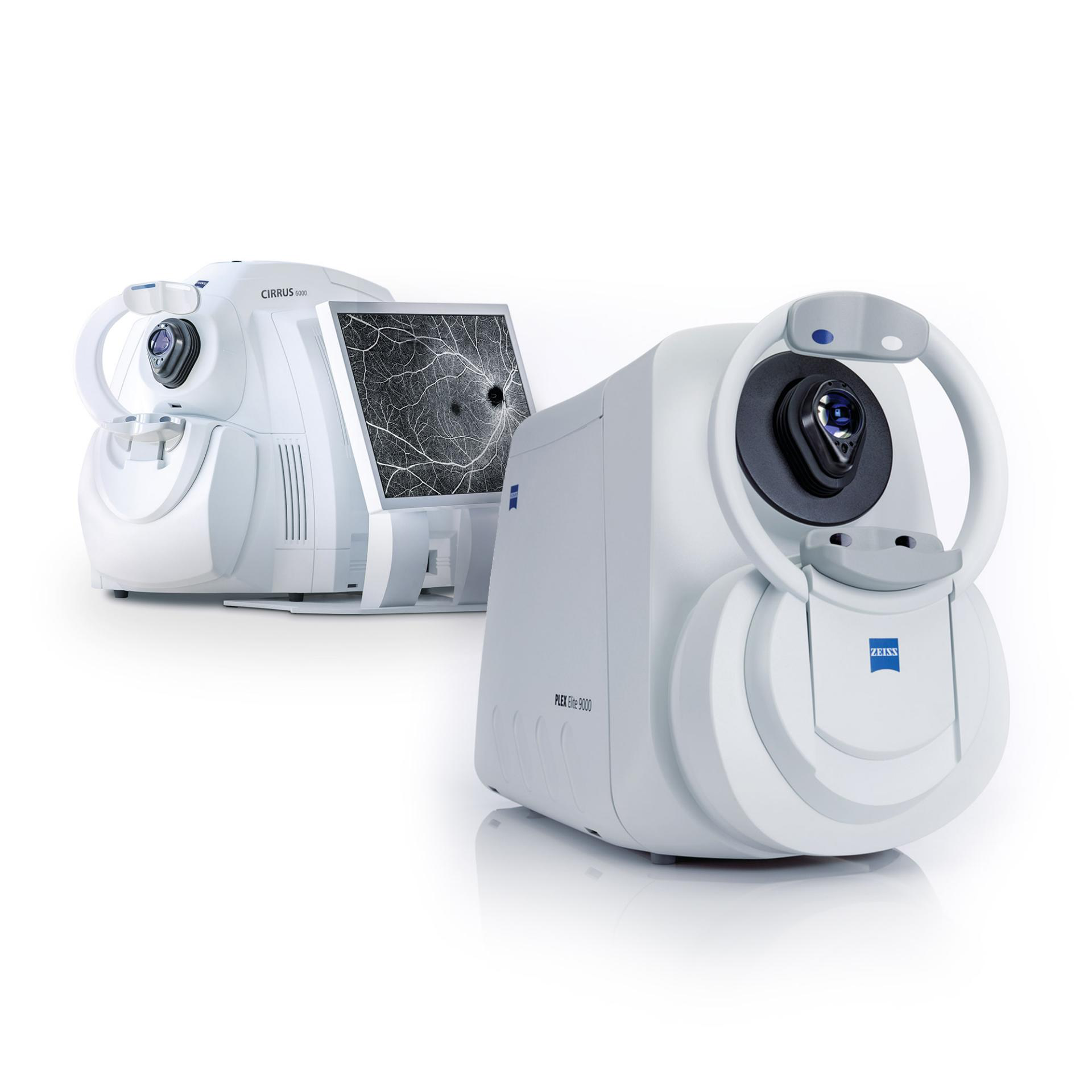
Optical Coherence Tomography (OCT)
Produces cross-sectional scans of the retina and macula.
Diabetic Retinopathy →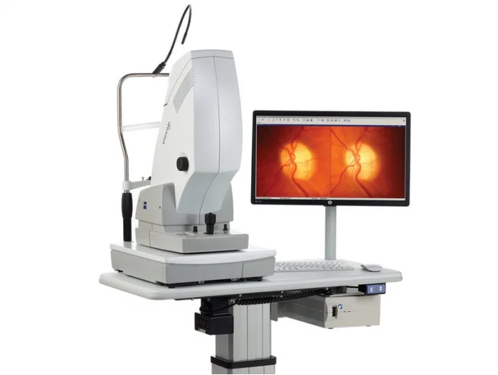
Retina
Fundus camera and angiography
Documents the retina and assesses retinal circulation.
Retinal Vein Occlusion →Patient reviews from Google
Verified public Google reviews for Perfect Vision Laser Eye Hospital – Dr. Virendra Laser Phaco Surgery Centre, Jaipur. Reviews are individual patient opinions and are not a guarantee of similar results.
“I was done with cataract surgery by virendra agrawal I m happy with his treatment as he given to me because I am diabetes also have retina problem but now I happy with his treatment”
“They say, god is around you! Well, we are so grateful, delighted to meet Dr Veerendra / Dr Anita and their entire staff. I feel blessed that they did everything to ensure to get properly diagnosed. Despite weekend and extremely busy schedules and Covid special time, they took utmost care and managed competing priorities. We are falling short of words but still trying our best to express gratitude. We are truly blessed & thankful for early diagnosis. Thank you so so much and we will do my best to follow your advice to the T. You guys are amazing and we surely recommend this place a modern temple to get your periodic proactive eye check ups or any other ophthalmic issues. Way to go!”
“The staff and doctors were very nice and there was not a very long wait time. Great organization and system in the hospital. All the eye tests were super easy to understand and were done efficiently. The doctors were very gentle and caring giving tissues whenever needed and checking if you are ok. I definitely would recommend this hospital for any eye problems or even just a checkup. My doctor was Dr. Neha Patak, she was extremely soft spoken and really caring. Salik was very gentle when lifting eyelids and that was very much appreciated.”
Relevant articles
Macular Hole — Jaipur FAQs
Related treatments
Retina & Uvea Overview
Comprehensive evaluation and treatment of retinal and uveal conditions.
Read moreDiabetic Retinopathy
Retinal damage caused by long-standing diabetes, often silent in early stages.
Read moreRetinal Detachment
A separation of the retina from its underlying layer, requiring urgent attention.
Read moreAge-Related Macular Degeneration
An age-related condition affecting the central retina.
Read moreDiscuss macular-hole treatment
Arrange OCT assessment and an individual discussion of observation or surgery.